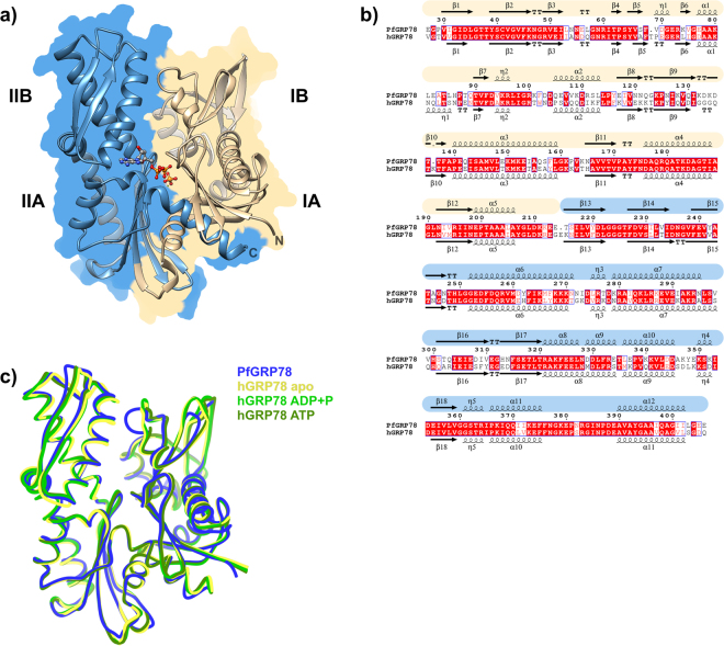
Figure 1.
Crystal structure of PfGRP78-NBD and comparison with the human GRP78 structures. (a) Cartoon representation of PfGRP78-NDB with each lobe colored differently (I – yellow and II – blue), with ADP and PO4 shown in ball and stick representation. (b) Sequence comparison between P. falciparum and human GRP78 NBDs. Secondary structure is shown above and below its corresponding sequence, and the malaria is color-coded according to the lobe organization as indicated before. (c) Structural overlay of malaria and human protein structures.

