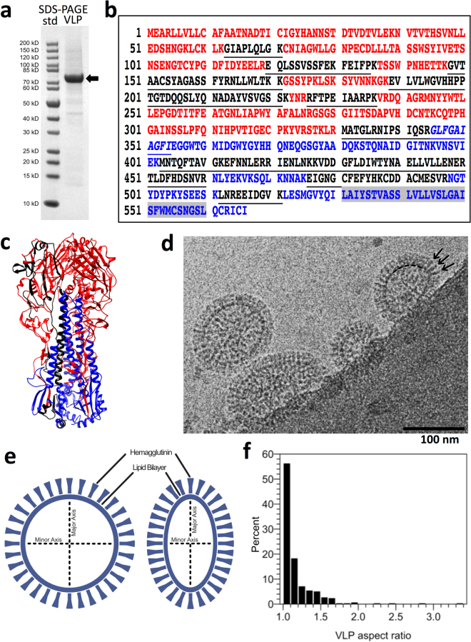
Figure 1.
Analysis of composition and organization of 1918 hemagglutinin (HA) H1 virus-like particles (VLPs) by peptide-finger printing and cryo-electron coupled with axial measurements. (a) SDS-PAGE analysis of VLPs under reducing conditions. Molecular weight standards (std) are in lane 1 and the VLP sample is in lane 2. An arrow denotes a band at an apparent molecular weight of about 75 kilodaltons. (b) Peptide finger printing of the major 75 kDa protein band of VLPs by mass spectrometry of tryptic peptides with database query of peptide profile matched to hemagglutinin sequence (A/Brevig Mission/1/1918 (H1N1)) by mass spectrometry with 35% coverage. The HA1 region sequence (M1-R344) is colored red with the HA2 region (G345-I566) in blue. The transmembrane region of HA2 is highlighted in grey. However, matched HA peptide regions are colored black within the sequence. (c) Peptides (black) mapped on the crystal structure of H1 HA from 1918 (PDB ID 1RD8) with HA1 and HA2 regions in red and blue, respectively. For clarity, the peptides are only mapped onto one HA1-HA2 protomer on the left-side. (d) Image by cryo-electron microscopy of a field of VLPs. Arrows denote some protruding spikes on the surface on a particle. The particles have oval-shaped outlined perimeters at the base of the protruding spikes. A portion of this outline in one particle is denoted by a hashed arc near the base of the indicated spikes (right particle). Scale bar, 100 nm. (e) Schematics of spherical and elongated VLP morphologies defined with a system of two axes as minor (shorter) and major (longer) that are used to determine aspect ratios and lengths. The dark peripheral outline and the spikes observed in cryo images are schematically assigned to the lipid membrane and hemagglutinin, respectively. (f) Distribution of VLP aspect ratio for a population of VLPs (N = 295). The aspect ratio is the ratio between major and minor axes as measured in x and y directions from cryo-EM images. Based on aspect ratios VLPs were assigned as spherical (1.0–1.1), near spherical (>1.1 and <1.5) and elongated (>1.5).

