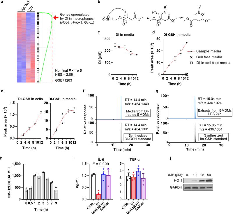
Extended Data Fig. 1. Detection of DI-GSH and Ita-GSH and electrophilic stress response.
a, Transcriptional comparison of KpCKO and wild-type BMDMs and enrichment of the DI gene signature. b, The reaction of DI with a thiol group in a Michael reaction. c, DI levels in media of BMDMs treated with DI for the indicated time, as determined by GC–MS. Mean of n = 2 cultures. d, Levels of the DI-GSH conjugate in the media of BMDMs treated with DI for the indicated time, as detected by LC–MS. Mean of n = 2 cultures. Data from Fig. 1e are overlaid with data for cell-free media. e, Levels of DI-GSH conjugate in BMDMs (left) and in their media (right) after treatment with 13C5-labelled DI for the indicated time, as detected by LC–MS. Mean of n = 2 cultures. f, g, Representative extracted ion chromatograms of DI-GSH detected in the media of BMDMs treated with DI for 6 h compared to the synthesized DI-GSH standard (f), and Ita-GSH detected in BMDMs stimulated with LPS for 24 h compared to the synthesized Ita-GSH standard (g). n = 10 technical replicates. h, Detection of reactive oxygen species in BV2 cells treated with DI for the indicated time, as determined by flow cytometry. Mean of n = 2 experiments. i, Cytokine production in BMDMs treated with DI in the presence of EtGSH and stimulated with LPS for 4 h, mean ± s.e.m., n = 3 experiments. j, Western blot of HO-1 expression in BMDMs treated with DMF. Representative of three experiments. For gel source data, see Supplementary Fig. 1. Statistical tests used were two-tailed t-tests.

