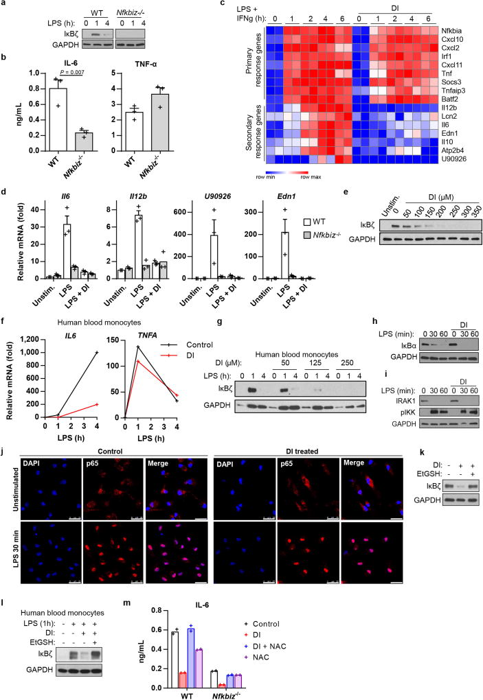
Extended Data Fig. 2. DI downregulates secondary transcriptional response to TLR stimulation.
a, Western blot of IκBζ expression in wild-type or Nfkbiz−/− BMDMs stimulated with LPS. b, Cytokine production in wild-type and Nfkbiz−/− BMDMs stimulated with LPS for 4 h, mean ± s.e.m., n = 3 experiments. c, RNA-seq analysis of BMDMs treated with DI and stimulated with LPS and IFNγ. d, mRNA expression show the induction of the indicated target genes in wild-type and Nfkbiz−/− BMDMs treated with DI and stimulated with LPS for 4 h, mean ± s.e.m., n = 3 experiments. e, Western blot of IκBζ expression in DI-treated BMDMs stimulated with LPS for 1 h. f, mRNA expression in human blood monocytes treated with DI and stimulated with LPS. g, Western blot of IκBζ expression in human blood monocytes treated with DI and stimulated with LPS. h, i, Western blot of IκBα (h) and IRAK1 expression and IKK phosphorylation (i) in BMDMs treated with DI and stimulated with LPS. j, p65 localization in DI-treated, LPS-stimulated BMDMs. Nuclei are stained with DAPI. Scale bars, 25 µm. Representative of two cultures. k, Western blot of IκBζ expression in BMDMs treated with DI in the presence of EtGSH and stimulated with LPS for 1 h. l, Western blot of IκBζ expression in human blood monocytes treated with DI in the presence of EtGSH and stimulated with LPS for 1 h. m, Cytokine production in wild-type or Nfkbiz−/− BMDMs treated with DI in the presence of NAC, stimulated with LPS for 4 h. Mean of n = 2 cultures. Representative data from two experiments (a), three experiments (e, h, i, k), three donors (f, g) and two donors (l). For gel source data, see Supplementary Fig. 1. Statistical tests used were two-tailed t-tests.

