Figure 1.

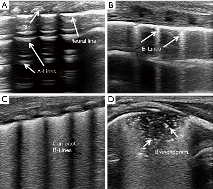
Ultrasound signs of the lungs. (A) A-line in a normal lung ultrasound image; (B) sonographic appearance of B-lines; (C) sonographic appearance of compact B-lines known as white lung; (D) subpleural lung consolidation with air bronchograms.

Ultrasound signs of the lungs. (A) A-line in a normal lung ultrasound image; (B) sonographic appearance of B-lines; (C) sonographic appearance of compact B-lines known as white lung; (D) subpleural lung consolidation with air bronchograms.