
FIGURE 3.
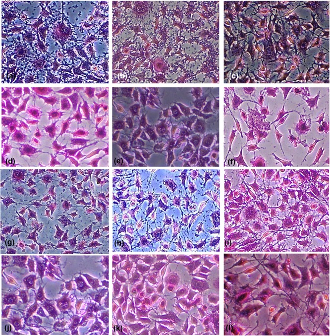
Light micrographs of Giemsa-stained untreated fibroblast cells showing adherence of (a) Enterococcus faecalis, (b) Streptococcus mutans, and (c) Candida albicans, to the cells a swell as the polystyrene of the microtitration plate. The light micrographs (d–f) show cells treated with Ch-NPs, (g–i) treated with Ag-NPs, (j–l) treated with O3-oil. Magnification 20×.