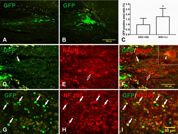
Fig. 5.
Differentiation of transplanted neural stem cells (NSCs) in tibial nerve in host spinal cord after spinal cord injury. (A–C) Transplanted NSCs survived well; (D) NSCs in TN + NSC + lithium chloride (LiCl) group sent out long neurites (arrow) and grew into host parenchyma; (E) host motoneurons (hollow arrow); and (F) merged picture. Upper right is magnified picture of dotted box in the middle. A contact of regenerated axon with host neuron can be seen (arrow head); (G) green fluorescent protein (GFP)-positive NSCs; (H) NF200-positive expression in cell body and neurites; (I) merged picture indicates double-labeled cells showing yellow color (arrow).

