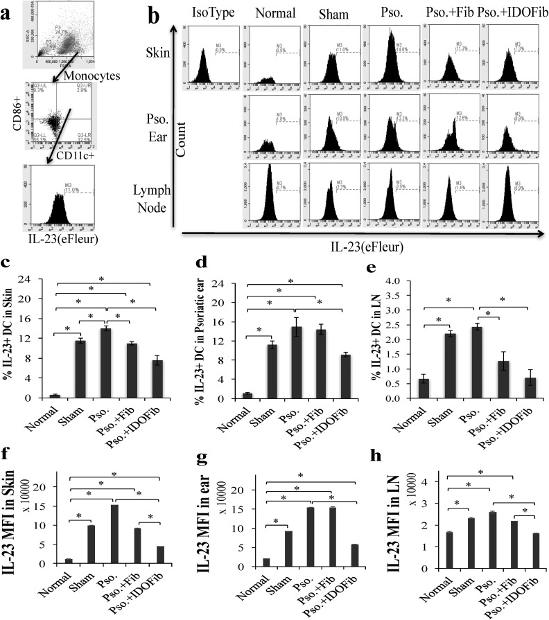
Fig. 8.
Flow cytometry analysis of the percentage of IL-23+ dendritic cells in skin, ear, and lymph nodes. Upon euthanizing mice on day 9, the dorsal skins, right ears, and lymph nodes were collected. After obtaining single-cell suspensions, cells were stimulated in Cell Stimulation Cocktail overnight. Dendritic cells were stained for their surface and intracellular markers in 1% fetal bovine serum + phosphate buffered saline–containing fluorescein isothiocyanate-conjugated anti-CD11c, phycoerythrin-conjugated anti-CD86, and eFleur-conjugated anti-IL-23. (a) Representative flow cytometry plots of gating. (b) Representative flow cytometry plots of IL-23+ CD11c + DCs in skin, ear, and lymph nodes, respectively. (c to e) Quantitative analysis of IL-23+ CD11c + DCs in skin, ear, and lymph nodes, respectively. (f to h) Mean fluorescence intensity (MFI) of IL-23 in skin, ear, and lymph nodes, respectively. Pso. ear, right ear which has received 5 mg imiquimod cream. The significant differences have been indicated by asterisks (*; P < 0.01; n = 3).

