Fig. 6.
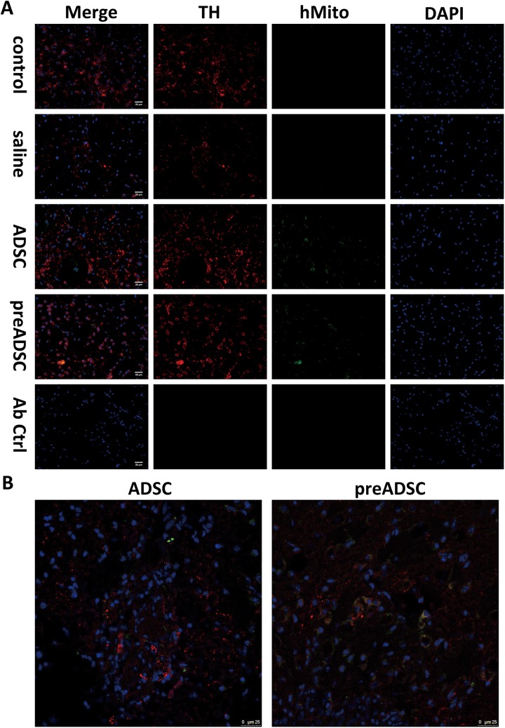
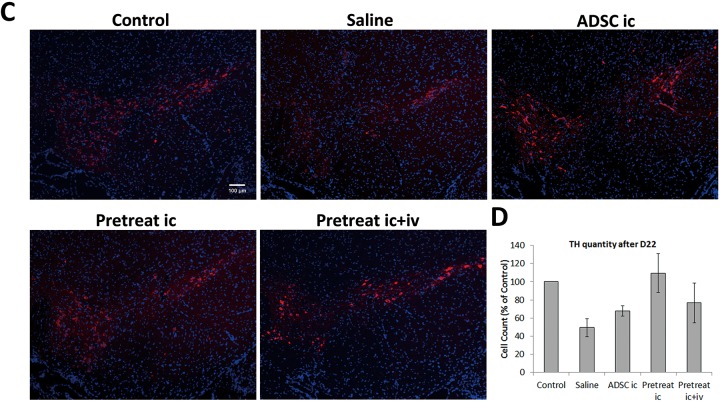
Number of tyrosine hydroxylase–expressing cells in mice transplanted with n-butylidenephthalide-pretreated adipose-derived stem cells (ADSCs). (A) Striatum of mice from control, saline, ADSC, preADSC groups were stained with tyrosine hydroxylase (red), human mitochondria marker (green), and 4′,6-diamidino-2-phenylindole (DAPI; blue), scale bar = 25 μm. (B) Colocalization of tyrosine hydroxylase (TH) and human mitochondria from (A) was confirmed by confocal microscopy. (C) The substantia nigra of mice from each group was stained for TH (red). DAPI (blue) was used for staining of nuclei. (D) Cells positive for both TH and DAPI were counted and presented as percentage versus control in the bar graph. Error bars represent means ± SDs (N = 3).


