Figure 2.

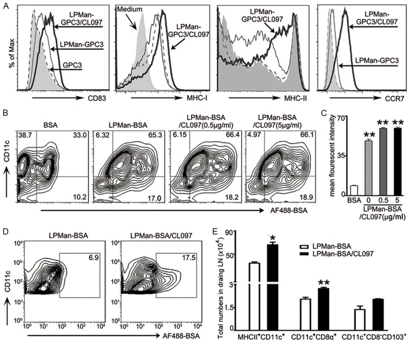
Effect of LPMan-GPC3/CL097 on migratory DCs and draining lymph nodes in vivo. A. FCM analysis of cell surface markers on BMDCs (3×10⁶/ml) by staining with antibody to CD83, MHC-I, MHC-II and CCR7 after they were stimulated for 24 h with LPMan-GPC3/CL097 containing 5 μg/ml GPC3 plus 0.5 μg/ml CL097. The same amount of LPMan-GPC3 containing 5 μg/ml GPC3, or free GPC3 (5 μg/ml) were used as control. Profiles show representative of three independent experiments. B, C. AF488-labeled BSA (BSA) was used as the model antigen and encapsulated with LPMan as performed as GPC3. BMDCs (3×10⁶/ml) were stimulated with LPMan-BSA/CL097 containing 5 μg/ml-BSA plus 0.5 μg/ml-CL097, or LPMan-BSA/CL097 containing 5 μg/ml BSA plus 5 μg/ml CL097, or LPMan-BSA containing 5 μg/ml BSA, or 5 μg/ml free BSA for 30 min. The percentage (B) and amount (C, as indicated by mean fluorescent intensity) of BSA in CD11c+ DCs was determined. **P<0.01 compared with the group treated with free BSA. D, E. Each mouse received 5 μg of AF-BSA in the form of LPMan-BSA (n=5) or received 5 μg of AF-BSA plus 5 μg of CL097 in the form of LPMan-BSA/CL097 via subcutaneous injection (n=5). raining lymph nodes were removed 48 h later. D. Profiles show the presence of AF-BSA in CD11c+ cells of the draining lymph nodes. E. Bars show the total numbers of MHCII+CD11c+ cells, CD11c+CD8α+ cells and CD11c+CD8α-CD103+ in draining lymph nodes of each mouse. Data are shown as mean ± SE, and un-paired t test was conducted between the two groups. *P<0.05; **P<0.01.
