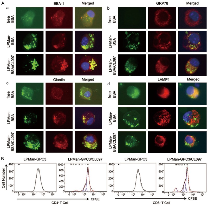
Figure 3.

Fate of internalized antigens in DCs delivered by mannosylated liposomes. A. AF488-labeled BSA (BSA, green) was used a model antigen. BMDCs were treated for 3 h with 5 μg/ml BSA alone, or LPMan-BSA containing 5 μg/ml BSA, or LPMan-BSA/CL097 containing 5 μg/ml-BSA plus 0.5 μg/ml-CL097. The cells were washed and placed on poly-L-lysine-coated slides and then were fixed with 2% paraformaldehyde for 10 min. The cells were stained with Cy3-labeled (red) antibodies of anti-EEA-1 (a), anti-Grp78 (b), anti-Giantin (c) or anti-LAMP1 (d). One representative experiment of five is shown. B. BMDCs were treated with LPMan-GPC3/CL097 containing 5 μg/ml GPC3 plus 0.5 μg/ml CL097, or same amount of LPMan-GPC3 containing 5 μg/ml GPC3 for 24 h. Untouched T cells were isolated from naïve mice and labeled with 5 μM CFSE. The two cell populations were co-cultured at the ratio of 1-BMDC to 20-T cells for 5 days. CD4+ T cell (left panel) and CD8+ T cell (right panel) proliferation levels were determined by FCM (one representative experiment out of three).
