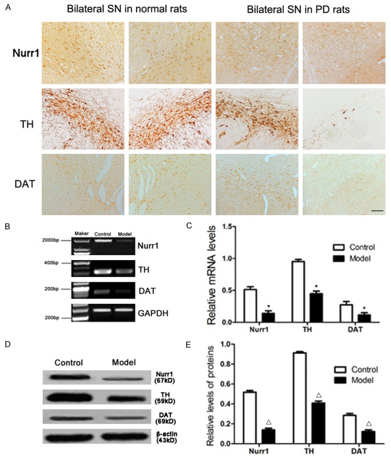
Figure 4.

Expression of Nurr1, TH and DAT in the substantia nigra of PD rats detected by immunohistochemistry, RT-PCR and Western blot. Microphotographs showed the Nurr1, TH and DAT immunoreactive neurons (A). Representative RT-PCR of Nurr1, TH and DAT in normal and PD rats. GAPDH was used as an internal control (B). The relative mRNA level of Nurr1, TH and DAT was analyzed by RT-PCR (C). Representative Western blot of Nurr1, TH and DAT protein in normal and PD rats. β-actin was used as an internal control (D). The relative intensity of Nurr1, TH and DAT protein was analyzed by Western blot (E). Data are presented as mean ± SEM. *P<0.01, Δ P<0.01 versus PD rat. The number of rats in each group was as follows: control (n = 3), model (n = 5). Scale bar = 50 μm.
