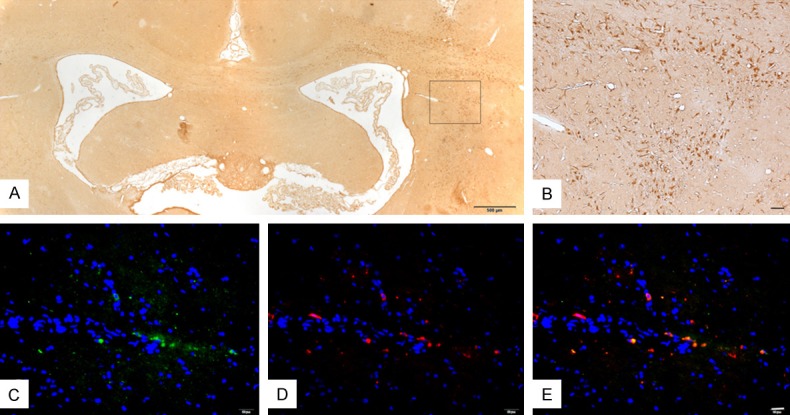
Figure 5.

Survival and migration and differentiation of implanted cells in the rat brain of the Lv-Nurr1-MSCs group four weeks after transplantation by immunohistochemical and double immunofluorescence staining. Lv-Nurr1-MSCs were injected into the striatum with microsyringe. The number of Nurr1-positive cells in the right striatum was significantly more than that in the left side, and cell migration was visible. Nurr1-positive cells were in the different part of the brain. (A). Nurr1-positive cells were fusiform or polygonal, and some like neurons. (B) is the enlarged view of (A) (B). Double immunofluorescence staining of TH (red) and Nurr1 (green) and nucleus (blue) (C-E). The number of rats in LV-Nurr1-MSCs group was five. Scale bar: (A) = 500 μm; (B) = 50 μm; (C-E) = 20 μm.
