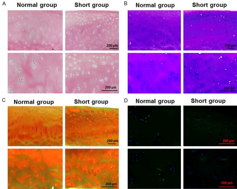
Figure 1.

Analysis of femoral cartilage morphology and Egr1 expression in the cartilage of patients. Microscopic appearance of the femoral head cartilage of patients suffering from necrosis of the femoral head was observed. Paraffin-embedded sections were stained with (A) H&E, (B) Toluidine blue or (C) Safranine-O. Magnification was 100× and 200×. (D) Representative sections from patient cartilage were probed for Egr1 by immunofluorescence. Magnification was 200× and 400× (Normal height patient N=5, shorter height patient N=5).
