Figure 6.

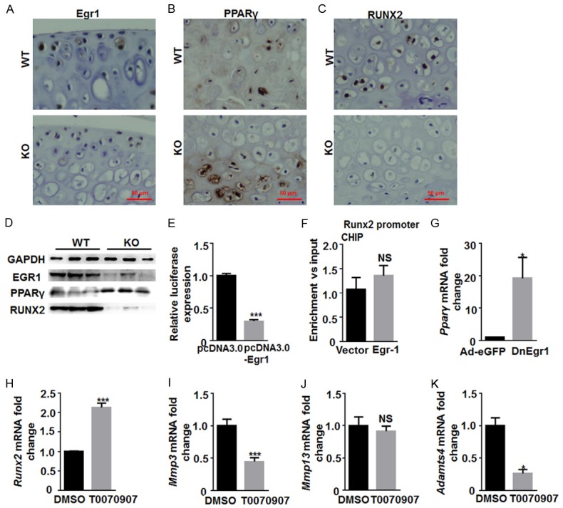
Egr1/PPARγ/RUNX2 signaling pathways participate in the regulation of the chondrocyte extracellular matrix. Representative images of the immunohistochemical staining for (A) Egr1, (B) PPARγ and (C) Runx2 in the Egr1 KO and WT mice cartilage. (D) Western blot was used to analyze the Egr1, PPARγ and Runx2 protein expression in the Egr1 KO and WT mice. GAPDH was used as control. (E) A luciferase reporter gene assay for the detection of Egr1 effect on Runx2. (F) Chromatin immunoprecipitation was performed in iMACs using antibodies of Egr1 to examine the direct interaction between Egr1 and Runx2. Precipitated DNA was analyzed by real-time PCR. (G) qRT-PCR analysis of PPARγ mRNA was performed after transfected DnEgr1 virus into iMACs. The mRNA expression of (H) Runx2, (I) Mmp3, (J) Mmp13 and (K) ADAMTS like 4 in iMACs were analyzed by qRT-PCR after treated with PPARγ inhibitors, T0070907 or DMSO. (*P<0.05; **P<0.01; ***P<0.001).
