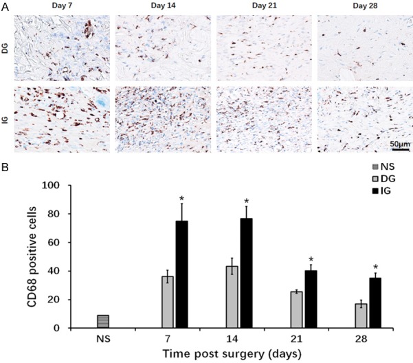
Figure 7.

Presentation of CD68 positive cells of the granulation tissue underneath the skin graft. A. CD68 staining of macrophage cells. Blue: cell nucleus; brown: CD68 positive cell. B. Quantification of macrophage cells at day 7, day 14, day 21 and day 28. *P<0.05. The IG group showed more severe inflammation throughout the wound healing process.
