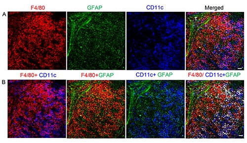
Figure 6.

Interaction of NMSCs and macrophages in red pulp of spleen from C57BL/6 mouse. Antibodies against F4/80 (red), CD11c (blue), and GFAP (green) label mainly macrophages, DCs, and NMSCs in the red pulp, respectively. b) Various combinations of channels in (a) are shown here. In the fourth image from left, the F4/80+CD11c+ DCs are marked in white after colocalization analysis. T, trabecula. Objective lens: 40x. Each image is a maximal intensity projection of a Z-stack. Stack size: 6.0 μm; optical slice interval: 0.50 μm. Scale bar: 20 μm.
