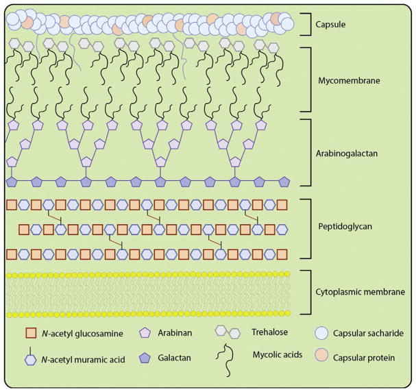
Fig. 1.
Simplistic cartoon depiction of the mycobacterial cell surface. The components of the cell envelope include: the cytoplasmic lipid bilayer, peptidoglycan, arabinogalactan, mycomembrane containing trehalose mycolic acids and capped with a capsule composed of polysaccharides, and capsular proteins.

