Figure 1.
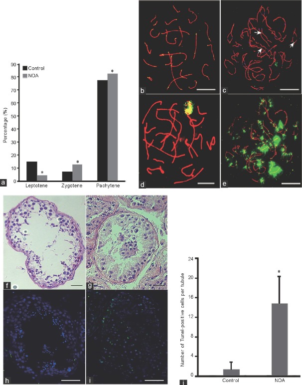
Analysis of meiotic progression and recombination in this NOA patient. (a) The percentages of leptotene, zygotene, and pachytene cells in the NOA patient and controls in meiosis prophase I. (b) Characteristic appearance of pachytene stages of germ cells from the controls, visualized using antibodies against synaptonemal complexes (red), MLH1 (green). (c) Abnormal recombination and γ-H2AX staining of pachytene stages from the NOA patient, MLH1 foci were observed in the chiasmata between homologous chromosomes (white arrow), visualized using antibodies against synaptonemal complexes (red), MLH1 (green). (d) γ-H2AX staining of pachytene cells from the controls, visualized using antibodies against γ-H2AX (green). (e) Amount of γ-H2AX foci occurred in the same cell with MLH1 in this NOA patient. (f) HE staining of testis tissue from the controls. (g) HE staining of testis tissue from the azoospermic man. (h) TUNEL staining of testis tissue from the controls. (i) TUNEL staining of testis tissue from the azoospermic man. (j) The average TUNEL-positive cell (green) number per seminiferous tubule of the patient and controls. Scale bars = 5 μm in b–e and 50 μm in f–i. *A significant difference with P < 0.05. NOA: nonobstructive azoospermia; HE: hematoxylin–eosin; MLH1: mutL homolg 1; TUNEL: terminal deoxynucleotidyl transferase dUTP nick end labeling; γ-H2AX: γ-H2A histone family, member X.

