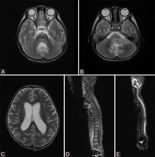
Figure 1(A-E).

(A-C) First MRI brain showed T2W hyperintense signal in anterior pons, medulla and bilateral cerebellar hemispheres. Obstructive hydrocephalus is evident. No abnormal signal is seen in bilateral cerebrum. (D and E) First MRI spine showed generalized swelling and increase in T1W hypointense and T2W hyperintense signal affecting the whole spinal cord
