Figure 2(A-D).

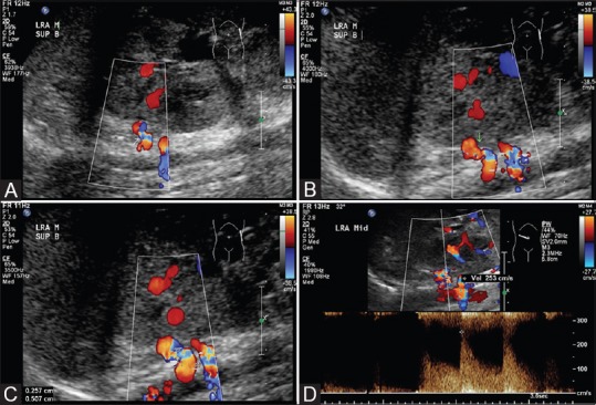
(Doppler USG study of renal arteries): (A-C) Doppler USG showing focal narrowing with aliasing (arrow) in superior branch of left renal artery. (D) Doppler USG showing elevated peak systolic velocity up to 253cm/s at the stenosis

(Doppler USG study of renal arteries): (A-C) Doppler USG showing focal narrowing with aliasing (arrow) in superior branch of left renal artery. (D) Doppler USG showing elevated peak systolic velocity up to 253cm/s at the stenosis