Figure 4(A-C).

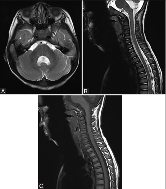
(A) Follow up MRI in 6 weeks showing resolution of abnormal signal and swelling in brainstem and cerebellum. (B and C) Follow up MRI spine in 6 weeks showing resolution of spinal cord abnormal signal and swelling

(A) Follow up MRI in 6 weeks showing resolution of abnormal signal and swelling in brainstem and cerebellum. (B and C) Follow up MRI spine in 6 weeks showing resolution of spinal cord abnormal signal and swelling