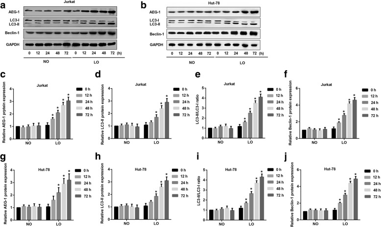
Fig. 2.
Effect of hypoxia on AEG-1 and autophagy markers in T-NHL cells. Hut-78 and Jurkat cells were incubated under normoxia or hypoxia for 0, 12, 24, 48 and 72 h before detection. a Expression of AEG-1, Beclin-1, LC3-I and LC3-II in Jurkat cells under normoxia and hypoxia environment via Western blot assays. b Expression of AEG-1, Beclin-1, LC3-I and LC3-II in Hut-78 cells under normoxia and hypoxia environment via Western blot assays. c-f Quantitative analysis of AEG-1, Beclin-1, LC3-I and LC3-II in Jurkat cells under normoxia and hypoxia. The expression of AEG-1 (p < 0.05), LC3-II (p < 0.05) and Beclin-1 (p < 0.05) was much higher in hypoxia than normoxia at 12, 24, 48 and 72 h. g-j Quantitative analysis of AEG-1, Beclin-1, LC3-I and LC3-II in Hut-78 cells under normoxia and hypoxia. The expression of AEG-1 (p < 0.05), LC3-II (p < 0.05) and Beclin-1 (p < 0.05) was much higher in hypoxia than normoxia at 12, 24, 48 and 72 h. NO: normoxia, NO: hypoxia. *p < 0.05, NO vs. LO

