Figure 3.
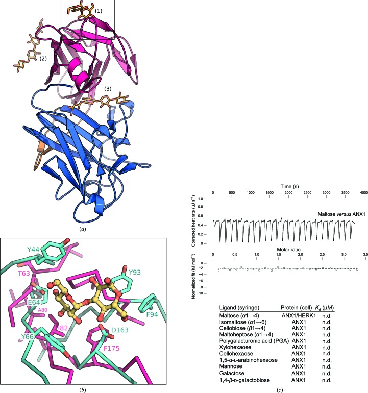
The malectin-like domains of ANX1 do not form canonical carbohydrate-binding sites. (a) Structural superposition of carbohydrate-binding sites from the malectin protein from X. laevis (PDB entry 2k46; Sainz-Polo et al., 2015 ▸) and CBM22 from P. barcinonensis (PDB entry 4xur; Montanier et al., 2009 ▸) onto ANX1 mal-N and mal-C (coloured as in Fig. 1 ▸). Carbohydrates are shown in bond representation (in yellow). (1) The nigerose-binding site of X. laevis malectin maps to the upper side of mal-N. (2) The binding surface of the xylotetraose in CBM22 is absent in mal-N; however, it maps to a potential binding cleft in ANX1 when superimposed on mal-C (3). (b) Close-up view of the nigerose-binding pocket of X. laevis malectin (shown in cyan) superimposed on ANX1 mal-N (magenta). The residues involved in nigerose binding are shown in cyan (in bond representation); the corresponding residues in ANX1 (magenta) are not conserved. (c) Isothermal titration calorimetry of d-maltose versus the ANX1 ectodomain; the table shows a summary for different carbohydrate polymers (K d, equilibrium dissociation constant; n.d., no detectable binding).

