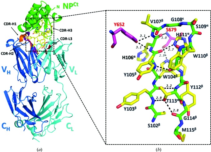
Figure 3.
Crystal structure of the BDBV NPCt–MJ20 complex. (a) Ribbon representation of the complex, with NP colored green and the MJ20 light chain and heavy chain colored cyan and blue, respectively. CDR3HC, which forms the majority of contacts with the antigen, is shown as yellow sticks. The variable domain (V) and the constant domain (C) as well as the four complementarity-defining regions (CDRs) are marked. (b) The secondary structure of the CDR3HC loop is shown as sticks with the main chain colored green and the side chains in yellow. The conformation of each residue is indicated in superscript. The CDR3HC loop adopts a β-hairpin stabilized by backbone–backbone/side-chain hydrogen bonds (marked as dashed lines with their lengths in Å). Two NPCt residues (Tyr652 and Ser679) that form five intermolecular hydrogen bonds to CDR3HC are shown as magenta sticks.

