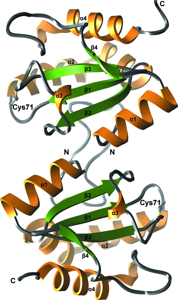
Figure 1.

Ribbon representation of the crystal structure of human AGR3. Two protein molecules of the asymmetric unit are represented. The core β-sheet is coloured green, α-helices orange and loop regions grey. The N- and C-termini and secondary-structure elements are labelled. The active-site Cys71 from the DCYQS motif is shown in ball-and-stick representation with C atoms coloured grey and the S atom in yellow.
