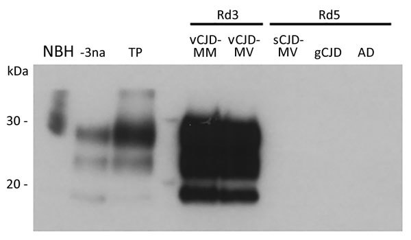
Figure.

Western blot analysis of vCJD prions obtained after amplification by protein misfolding cyclic amplification (PMCA) of cerebrospinal fluid (CSF) from 2 patients with vCJD (MM and MV) and 3 control patients and a crude reference brain homogenate from a vCJD patient (National Institute for Biological Standards and Control [Ridge, UK] no. NHBY0/0003). Abnormal prion protein patterns were assessed by using antibody 3F4 after digestion of samples with proteinase K. A total of 20 μL of each sample was subjected to electrophoresis on a 12% polyacrylamide gel. Lane NBH, negative control brain homogenate from a person without CJD and no digestion with proteinase K (National Institute for Biological Standards and Control no. NHBZ0/0005); lane -3na, Western blot control (10−3 dilution of vCJD reference brain sample without amplification); lane TP, positive control for amplification (10−6 dilution of vCJD reference brain sample after 1 round of PMCA); lane vCJD-MM, CSF from a patient with MM vCJD after 3 rounds of PMCA; lane vCJD-MV, CSF from a patient with MV vCJD after 3 rounds of PMCA; lane sCJD-MV, CSF from a patient with MV sCJD after 5 rounds of PMCA; lane gCJD, CSF from a patient with gCJD after 5 rounds of PMCA; lane AD, CSF from a patient with Alzheimer’s disease after 5 rounds of PMCA. CJD, Creutzfeldt-Jakob disease; gCJD, genetic CJD; MM, methionine homozygous; MV, methionine/valine heterozygous; Rd, round (1 round indicates 80 cycles of PMCA); sCJD, sporadic CJD; vCJD, variant CJD.
