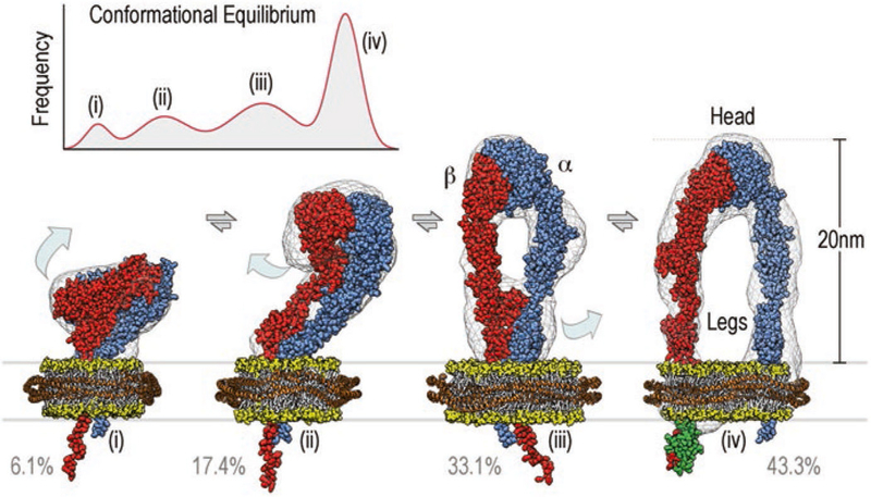
Fig. 12.1.

Integrin αIIbβ3 exists in a continuous conformational equilibrium centered around four main conformational states. The height of the peaks is given by the percentage of particles assigned to a given conformation, the width was estimated from the structural variability within each group. The equilibrium in the presence of the extracellular ligand RGD, cytosolic binding partner talin head, and lipid bilayers is depicted in the top left corner and centers around the four conformations shown in the lower part of the figure. Space-filling atomic models of αIIbβ3 integrin (α-subunit in blue, β-subunit in red) embedded in nanodiscs (lipid head groups in yellow, belt protein in orange) are shown fitted into their respective three-dimensional reconstructions (grey wire representation). Required structural transitions are depicted as light blue arrows. In the three-dimensional reconstruction of the upright state (iv) density for bound talin head (F3 domain shown in green) was evident
