Image 1.
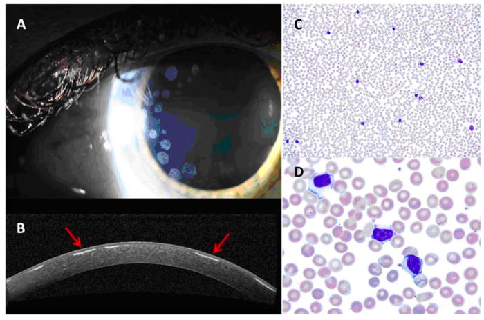
Slit lamp examination showing multiple, circular, peripherally located anterior corneal stromal deposits (A). Optical coherence tomography of the cornea shows deposits (arrows) with intervening clear spaces (B). Peripheral blood smear at 20× (C) and 100× (D), showing a mild lymphocytosis composed of small- to medium-sized mature lymphocytes with occasional smudge cells.

