Figure 1.
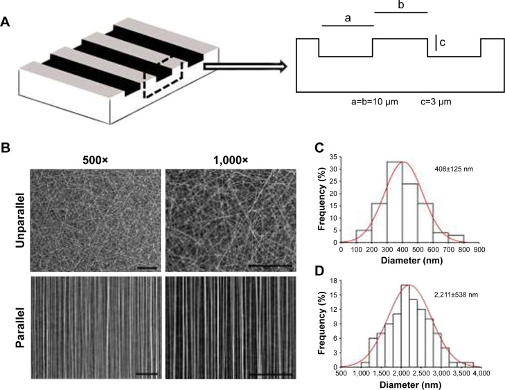
Characterization of nanoscaled and microscaled parallel topography. (A) The design of parallel microgrooved silicone membrane with groove width of 10 µm and groove depth of 3 µm. (B) SEM image of GT/PCL nanofibers showed unparallel and parallel patterns of electrospun nanofibers (500× and 1,000×, bar =100 µm). (C) The diameter distribution of unparallel nanofibers. (D) The diameter distribution of parallel nanofibers.
Abbreviations: GT, gelatin; PCL, poly(ε-caprolactone); SEM, scanning electron microscope.

