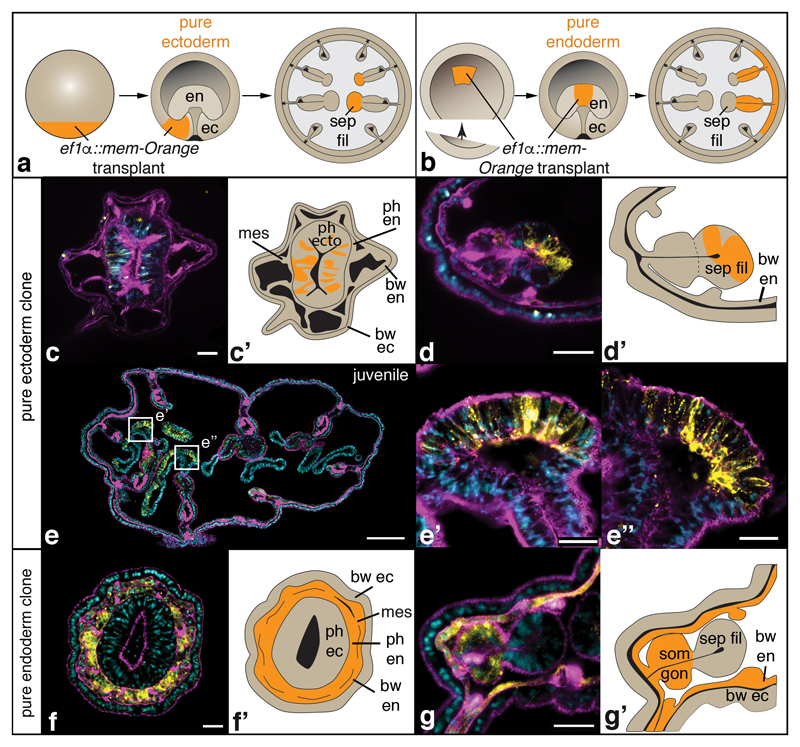
Figure 2. Fate mapping reveals an ectodermal origin of the pharynx and septal filaments.
Schematized transplantation experiment and fate map (a, b), single confocal images (c, d, e-e”, f, g) and schematics (c’, d’, f’, g’) of cryo-sectioned animals containing transplanted tissue (c-g’). Samples are exemplary primary polyps (c-d’, f-g’) or a juvenile polyp (e-e”) with pure ectodermal (c-e”) or pure endodermal (f-g”) integration of transplanted, mem-Orange-positive donor tissue. A detailed description of the transplantation techniques and results is provided in Supplementary Figure 1. Colours in confocal pictures: yellow: mem-Orange staining; magenta: F-actin staining; blue: nuclear staining (DAPI). bw: body wall; ec: ectoderm; en: endoderm; mes: mesentery; ph: pharynx; sep fil: septal filament; som gon: somatic gonad. Scale bars in c, d, f, g: 20μm; in e: 200μm; in e’, e”: 25μm.

