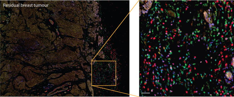
Figure 1.

Immune infiltration in a human breast carcinoma. Triple negative breast cancer residual tumour section shows infiltration of different immune cell populations in tumour stroma: CD4+ and CD8+ T cells, CD20 B cells FoxP3+ activated lymphocytes and CD68+ macrophages (CD4 (green), CD8 (red), CD20 (yellow), FoxP3 (magenta), CD68 (cyan), nuclei (blue)). The section was stained with the OPAL-7 Solid Immunology Kit and imaged on a Vectra Quantitative Pathology Workstation. Scale bar: 20μm.
