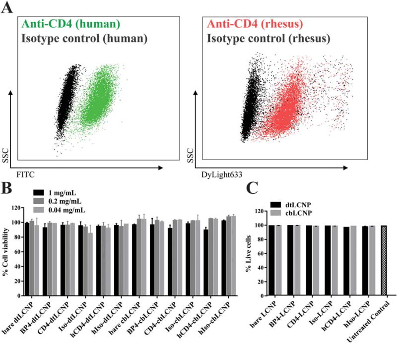
FIGURE 4.
(A) Flow cytometry dot plot analysis of CD4 staining on a 174×CEM human T cell line. Left: 174×CEM cells treated with FITC antihuman CD4 antibody (green) or FITC isotype IgG antibody (black). Right: 174×CEM cells treated with DyLight633 labeled rhesus recombinant anti-CD4 antibody (red) or DyLight633 labeled rhesus recombinant isotype IgG antibody (black). (B) dtLCNP or cbLCNP conjugated with different CD4 binding ligands showed no cytotoxicity to 174×CEM cells after treating cells with up to 1 mg/mL LCNPs for 24 h, measured by Cell Titer-Blue. Untreated cells were used as 100% viability control. (C) Flow cytometry analysis of live/dead staining showed that all LCNP formulations had no cytotoxicity to pigtail macaque PBMCs after incubaiton with 0.5 mg/mL LCNPs for 24 h. Data represents mean ± SD, n = 3.

