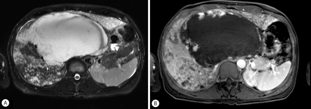
Figure 3.
Liver MRI image obtained on June 3, 2015. T2-weighted MRI image shows the enlarged huge mass with a high signal intensity involving segments 1, 4, 7 and 8 of the liver. The extents of disseminated small enhanced lesions are increased (A: T2-weighted image, B: contrast-enhanced T1-weighted image).

