
Figure 2.
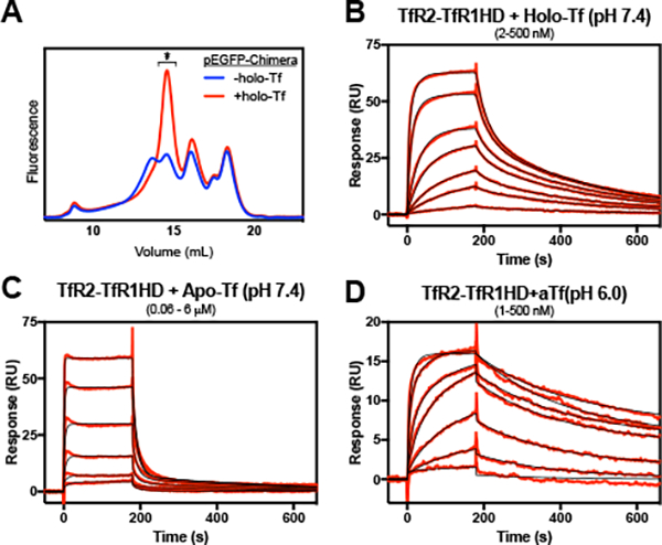
The interaction of a TfR2-TfR1 helical domain chimera with holo- and apo-Tf. The chimera receptor was solubilized in the absence or presence of 10 μM holo-Tf and applied to a Superose 6 column with in-line fluorometer (A). The receptor peak fraction from the +holo-Tf run (*) was immobilized to the SPR chip surface to a density of ~200 RU’s and treated with various concentrations of Tf. Concentrations of holo- or apo-Tf injected were the following: (B) 2 – 500 nM, with a 2.5-fold dilution series; (C) 0.06 – 6 μΜ, with a 2.5-fold dilution series; (D) 1 – 500 nM, with a 2.8-fold dilution series. Reference cell-subtracted binding response data is indicated in red, and heterogenous model fits are shown in black. Extrapolated kinetic constants from the model fits are shown in Table 1.