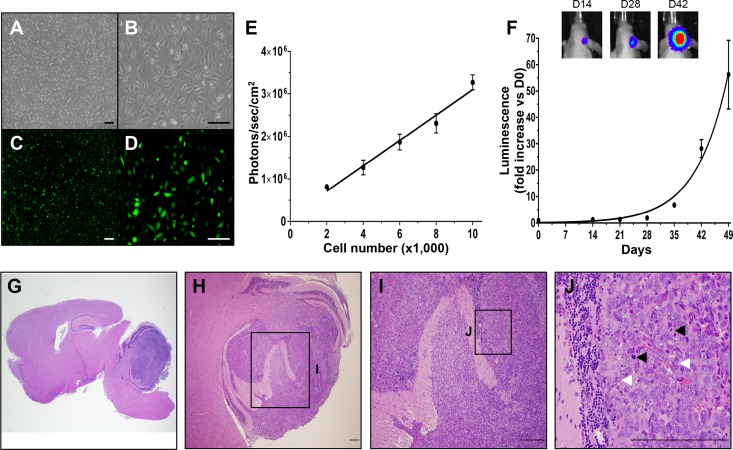
Fig 1. Daoy human MB cells engineered to express GFP and firefly luciferase form orthotopic xenografts in vivo.
Cultured Daoy human MB cells transduced with lentiviral vectors encoding GFP-FLuc (Daoy-GFPFL) express GFP and luciferase in vitro as determined by white light (A, B) and fluorescence imaging (C, D). Daoy-GFPFL cell number showed linear correlation with Fluc activity (R2 = 0.978, P = 0.001, E). Daoy-GFPFL xenografts showed exponential growth with a doubling time of 5.6 days in vivo measured by bioluminescence imaging. Representative bioluminescence images are shown for days 14, 24 and 42 after injection of Daoy-GFPFL cells (F). Representative hematoxylin and eosin staining (G-J) of brain sections show large intra-cerebellar tumors 63 days after injection of Daoy-GFPFL cells. Tumors showed histopathological features of MB, including frequent cell wrapping (white arrowheads) and heightened level of mitosis (black arrowheads). Original magnifications: 15X (G), 40X (A, C), 100X (B, D), 200X (H), 400X (I), 600X (J). Scale bars, 100 μm (A-D) and 200 μm (G-J). n = 5.

