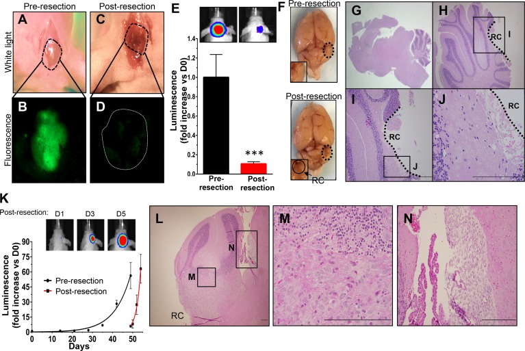
Fig 2. Fluorescence-guided microsurgical resection reduces volumes of orthotopic Daoy-GFPFL xenografts that redevelop.
A scalp incision was made 49 days after injection of Daoy-GFPFL cells and craniotomy (A). Fluorescence was used to visualize the underlying GFP+ tumors pre resection (B). Fluorescence-guided microsurgery significantly reduced tumor burden as determined by intra-operative imaging (C, D). Post-operative bioluminescence imaging showed a >92% reduction in mean tumor burden (***P = 0.009, E). Representative white light and bioluminescence images pre- and post-resection are shown (E,F). Representative H&E images (G-J) of brain sections taken immediately post-resection did not show apparent residual tumor within the resection cavity. Recurrent Daoy-GFPFL xenografts (K) grew faster than pre-resection counterparts (data from Fig 1F), with doubling times of 1.5 vs. 5.6 days, respectively (P = 0.0003). Representative BLI images are shown for days 1, 3, and 5 post-surgery. H&E images (L-N) of brain sections shows recurrent tumor near the resection cavity and cancer cells disseminating through the cerebral spinal fluid (CSF). Original magnifications: 15X (G), 200X (L, H), 400X (I), 600X (J, M, N). Scale bars, 200 μm. RC, Resection Cavity. n = 5 in each group.

