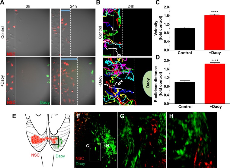
Fig 3. NSCs migrate towards human MB in vitro and in vivo.
Migration of NSC-mcF cells (red) the presence (+Daoy) or absence (control) of MB cells (Daoy-GFPFL cells, green) was monitored over 24h (A). Time-lapse images were captured every 20 mins and used to construct single cell tracings (B). Quantitative analysis of single cell tracings demonstrated the presence of Daoy-GFPFL increases the migratory velocity of NSCs (1.61-fold; ****P<0.0001, C), and euclidean distance traveled (1.8-fold; P<0.0001, D). Illustration of NSC and Daoy-GFPFL cells implanted in opposite cerebellar hemispheres (E). Fluorescent images (F-H) 21 days after stem cell implantation shows NSCs co-localize with Daoy-GFPFL tumor cells. Original magnifications: 100X (A, B,), 200X (F, G). n = 4 in panel E.

