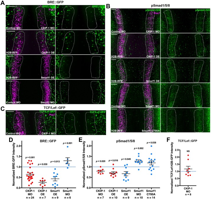
Fig 4. CKIP-1 and Smurf1 regulate BMP signaling and activation of Smad1/5/8.
(A) A BRE::GFP reporter construct was coelectroporated with indicated MOs or OE constructs. Embryos then were immunostained at HH7 for Pax7 and GFP. (B) Gastrulating embryos were electroporated with the indicated reagents and then immunostained at HH7 for Pax7 and activated pSmad1/5/8. (C) Embryos were electroporated with a TCF/Lef-driven H2B-GFP reporter construct along with control and CKIP-1 MOs and then immunostained for Pax7 and GFP at HH7. Scale bars represent 100 μm. (D-F) Fluorescence intensity for BRE::GFP (D), pSmad1/5/8 (E), and TCF/Lef::GFP (F) was quantified within the NPB (dashed white lines), normalized to the control side, and displayed with mean ± SEM. Underlying data can be found in S1 Data. P values from two-tailed Student t test. BMP, bone morphogenetic protein; BRE::GFP, BMP responsive element–driven GFP; CKIP-1, casein kinase interacting protein 1; GFP, green fluorescent protein; H2B, histone 2B; HH, Hamburger-Hamilton stage; MO, morpholino oligonucleotide; OE, overexpression; NPB, neural plate border; NS, not significant; OE, overexpression; Pax7, paired box 7; pSmad1/5/8; phospho-Smads 1/5/8; RFP, red fluorescent protein; Smurf1, Smad ubiquitin regulatory factor 1; TCF/Lef, T-cell factor/lymphoid enhancer factor.

