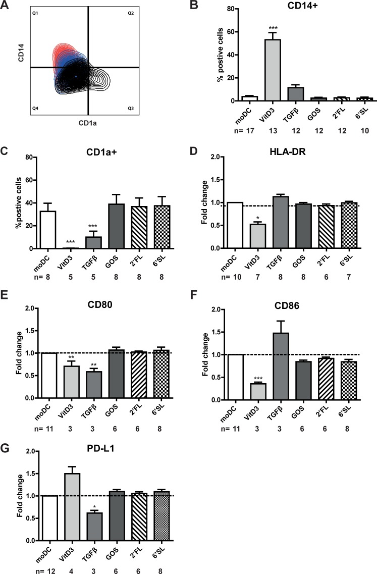
Fig 1. TGFβ and VitD3 induce phenotypic distinct DCs.
CD14+ monocytes were cultured in the presence of IL-4 and GM-CSF for six days in the presence or absence of breast milk components. Surface marker expression was measured by flow cytometry. A) A multi-colour overlay of CD14 expression versus CD1a expression on moDC (black), TGFβDC (blue) or VitD3 (red) of one representative donor is shown. The percentage of B) CD14+ and C) CD1a+ DC and relative surface marker expression of D) HLA-DR, E) CD80, F) CD86 or G) PD-L1 on immature DC differentiated in the presence of TGFβ, VitD3, 6’SL, 2’FL or GOS was shown. Relative fold change was calculated by dividing the MFI (median fluorescence intensity) of DC differentiated in the presence of a breast milk component/MFI of moDC of each respective donor.

