Figure 2.
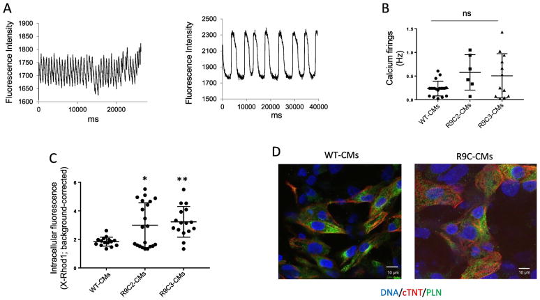
R9C hiPSC-CMs show abnormal calcium handling without PLN relocalization. (A) Two representative examples of calcium transients elicited in R9C hiPSC-CMs loaded with X-Rhod1 at day 40 of differentiation (paced at 0.25 Hz). (B) Total quantitation of calcium transients elicited of R9C hiPSC-CMs (day 40) compared to isogenic wild-type hiPSC-CMs paced at 0.25 Hz. Samples that captured correctly at 0.25 Hz are shown as a single point. (C) Diastolic calcium concentration of wild-type and R9C single hiPSC-CMs. (N=12–20). (D) Representative immunofluorescence images showing the intracellular protein distribution of phospholamban (PLN) and cardiac troponin T (cTNT) in wild-type and R9C hiPSC-CMs at day 35. Nuclei were counterstained with DAPI.

