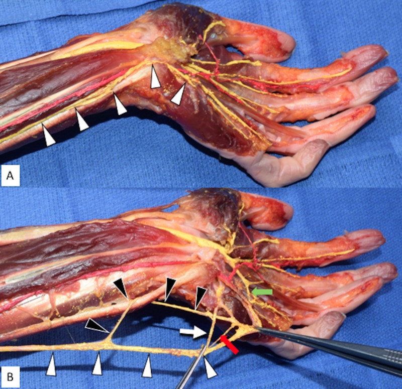
Figure 1. Left Anterior Forearm and Hand Dissection.
Figure 1A: Left palmar surface of the left hand and distal anterior forearm displaying the ulnar nerve in its anatomical position (arrowheads).
Figure 1B: Figure 1A with the ulnar nerve main trunk rotated 180 degrees and then retracted medially. Note the variant loop formed around the flexor carpi ulnaris. White arrowheads: ulnar nerve main trunk; Black arrowheads: variant loop around the distal tendon of the flexor carpi ulnaris; White arrow: deep branch of the ulnar nerve; Superficial branch of the ulnar nerve retracted at right forceps; Red arrow: additional nervous interconnection; Green arrow: Riché-Cannieu anastomosis between the superficial ulnar nerve branch and median nerve in the hand.

