
Figure 1.
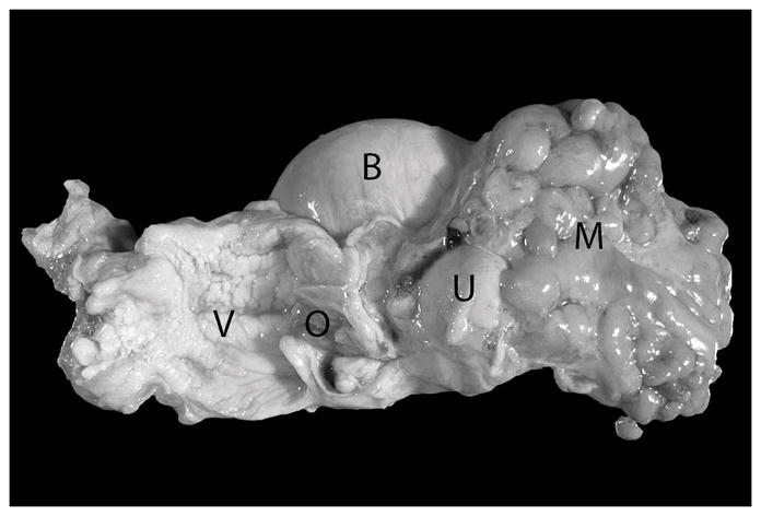
Vagina (opened – V), uterine isthmus with external orifice (opened – O), uterine body (U), and urinary bladder of a 20 year old Japanese macaque (Macaca fuscata). An approximately 8 cm in diameter firm mass encompassing the uterine horns and ovaries and partially incorporating the uterine body is present (M). Mesometrial and serosal adipose tissue is broadly adherent to this mass.