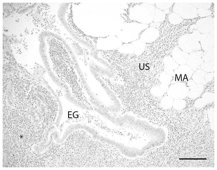
Figure 2.

The mass consisted histologically of coalescing nodules composed of well differentiated endometrial glands (EG) with adjacent uterine stromal tissue (US) infiltrating into the serosal and mesometrial adipose tissue (MA). Lymphocytes and plasma cells and occasional neutrophilic granulocytes are present in the tissue surrounding the ectopic uterine glands (asterisk). A mild cellular exudate composed of neutrophilic granulocytes is present within the lumen of the glands. (H&E stain; bar = 200 microns)
