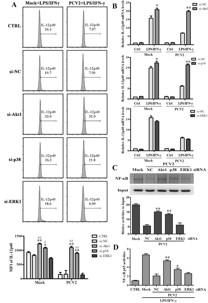
Figure 5. PCV2 infection activates PI3K/Akt and p38 MAPK signaling pathways to suppress IL-12p40 expression in transcriptional level.

The specific siRNAs of Akt1, p38 MAPK, ERK1, or negative control siRNA were transfected into PAMs for 24 h. Then the cells were infected by mock or PCV2 and followed by LPS/IFN-γ stimulation. The IL-12p40 expression were detected by flow cytometry (A) and qPCR (B). And the binding activities of NF-κB p65 to il12p40 promoter were measured using ChIP assay (C). (D) The NF-κB p65 activity was measured by Dual-Luciferase reporter assays. *P < 0.05, **P < 0.01 versus negative control siRNA transfected PAMs (si-NC) in same infection or Mock. #P < 0.05, ##P < 0.01 versus control PAMs (CTRL) in same infection or Mock.
