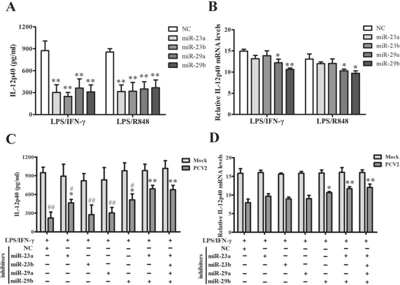
Figure 8. miR-23a and miR-29b play critical roles in post-transcriptional suppression of IL-12p40 expression in PCV2 infected cells.

(A, B) The miR-23a, miR-23b, miR-29a, and miR-29b mimics were transfected into PAMs for 24 h, then the cells were stimulated by LPS/IFN-γ or LPS/R848 for another 6 h or 24 h. The expression of IL-12p40 were measured by ELISA and qPCR. (C, D) The specific inhibitors of miR-23a, miR-23b, miR-29a, and miR-29b were transfected into PAMs separately or combined, then the cells were infected by 1 MOI PCV2 for 24 h. And the cells were further stimulated by LPS/IFN-γ for 6 h or 24 h. The expression of IL-12p40 were measured by ELISA and qPCR. (A, B) *P < 0.05, **P < 0.01 versus negative control mimic transfected cells. (C, D) *P < 0.05, **P < 0.01 versus negative control inhibitor transfected PCV2-infected cells. #P < 0.05, ##P < 0.01 versus the PCV2-infcted cells transfected with the mix of all 4 miRNA inhibitors.
