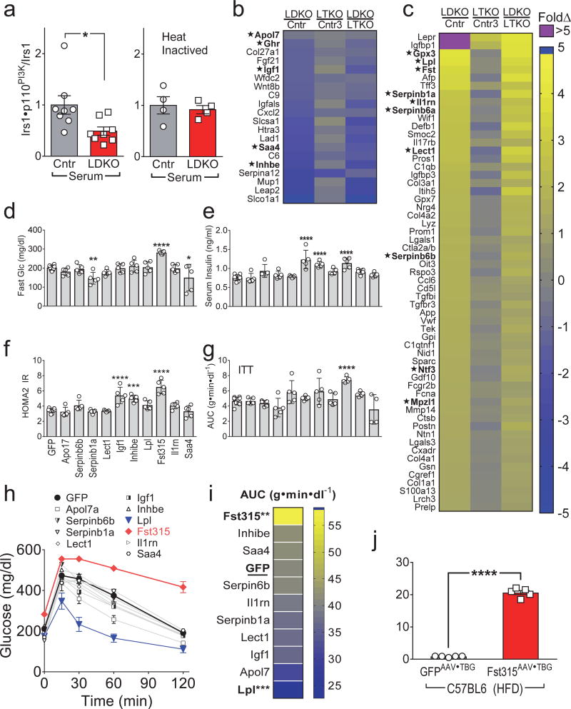
Figure 2. Identification of Fst315 as a hepatic FoxO1-regulated hepatokine regulating systemic glucose homeostasis.
(a) Insulin-stimulated Irs1•p110PI3K complex formation in 3T3-L1 adipocytes cultured with serum from overnight fasted four-month old LDKO (vs Cntr) mice without (left, n = 8) or with heat inactivation of the serum (56°C for 45 min.) (right, n = 4). (b,c) Identified genes encoding putative hepatic secreted proteins that decreased (b) or increased (c) >3-fold in LDKO mice (Benjamini-Hochberg FDR<0.05). Several of these secreted proteins seemed likely to have effects upon peripheral metabolism (marked as *) were selected for functional screening in mice. (d–i) Five-week old C57BL6 mice were challenged with high fat diet (45% fat) for 2 months, then infected with GFPAAV•TBG or hepatokineAAV•TBG (2×10¹¹ Genome Copy (GC)/mouse) encoding the indicated genes: fasting blood glucose (d) and fasting serum insulin (e) were measured one week after infection and used to calculate (f) HOMA2 IR as a measure of systemic insulin resistance (n = 5–10). (g) Insulin tolerance tests (ITT) were performed four weeks after infection with GFPAAV•TBG or hepatokineAAV•TBG and summarized by the areas under glucose curves (AUC) (n = 4–10). (h,i) Glucose tolerance tests were performed one week after AAV infection (h) and summarized (i) by the areas under glucose curves (AUC) (n = 4–10). (j) Fasting serum Fst levels measured four weeks after infection of HFD-fed C57BL6 mice with Fst315AAV•TBG or GFPAAV•TBG (n = 5). Data were analyzed by two-way ANOVA (h), one-way ANOVA (d,e,f,g,i) and unpaired Student’s t-test (a, j). Data are reported as the mean±SEM. *P<0.05; **P<0.01; ***P<0.001; ****P<0.0001.

