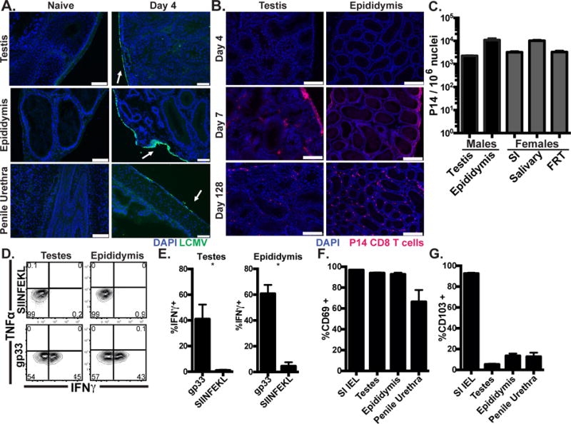
FIGURE 1. Resident-phenotype memory CD8 T cells persist in male genital tract.

(A) C57BL/6 mice were infected with LCMV i.p. and stained with anti-LCMV nucleoprotein antibody (Green), and DAPI (Blue). Scale bar=100μm. n=8 from 3 experiments. (B) Representative images 4, 7 or 128 days after Thy1.1+ P14 cell transfer and LCMV infection, Thy1.1+ P14 (red), DAPI (Blue), scale bar=250μm, n=6-8 from 2 or 3 experiments per time point. (C) Enumeration of Thy1.1+ P14 in tissues from male (black) or age-matched female (grey) mice 115-160 days after LCMV infection, n=6, representative of 4 experiments. (D-E) 120-160 days after LCMV infection, 50μg of gp33 (cognate) or SIINFEKL (irrelevant) peptide was transurethrally instilled. 12h later, P14 were isolated and cytokine expressing cells were assessed by flow cytometry. n=6, representative of 3 experiments. (F-G) CD69 and CD103 expression on P14 memory cells, n=12 from 3 experiments. Graphs show mean and SEM, *p 0.0357, Mann-Whitney.
