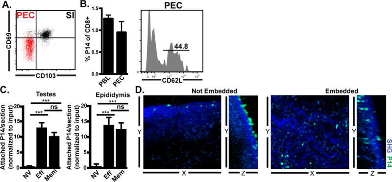
FIGURE 3. Recirculation through peritoneal cavity by memory CD8 T cells allows adherence and transcapsular migration to infected tissues.

(A) CD69 and CD103 on gated memory P14 CD8 T cells isolated from small intestine epithelium (SI, black) or peritoneal exudate cells (PEC, red). (B) Memory P14 CD8 T cells isolated from lymphoid organs were transferred i.v. into naïve mice. Donor cells were assessed in recipient blood (PBL) and peritoneal lavage (PEC) 18h later. (C) Naïve (NV), effector (Eff, 6 days after LCMV infection) or memory (Mem, 30 days after LCMV infection) congenically distinct P14 populations were isolated from spleen and co-injected i.p. (4-7.5×10⁶ of each cell type) into day 6 LCMV recipients. 3h later, attached P14 were quantified by immunofluorescence. Graph shows mean and SEM and results of Wilcoxon matched pairs statistical analysis, ***p 0.0005, ***p 0.001. (D) Memory P14 CD8 T cells were co-cultured for 2-3h with MGT freshly isolated from LCMV D6 mice. Live imaging was captured by 2-photon microscopy (representative still images shown). n=3 from 2 separate experiments.
