
Figure 3. The opposing effects of IL-36RA and IL-1Ra down-regulation on the outcome of Pa keratitis.
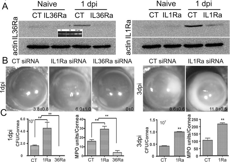
Mice were subconjunctivally injected with IL-1Ra, IL-36Ra specific, or the control, non-specific siRNA (50 picomoles in 5ul RNase free water) twice in two days. Corneas were inoculated with Pa (ATCC 19660, 1.0×10⁴ CFU), 6h after the second siRNA injection. (A) Western blotting assessing the efficacy of siRNA mediated knockdown of IL-1RA and IL-36RA in the cornea in response to Pa infection at 1 dpi with non-inoculated corneas (Naive) as the controls; β-actin serves as the loading control. CT: control, non-specific siRNA. Insert shows RT-PCR showing an increase in IL-36Ra levels at 1 dpi, compared with 0 and 18 hpi. (B) Mice were monitored and photographed daily up to 3 dpi, showing IL-36Ra- and IL-1Ra for 1 dpi and IL-1Ra for 3 dpi in-siRNA treated corneas. The numbers within each eye micrographs are the clinical scores assigned and presented as median + interquartile range. (C) At 1 dpi or 3 dpi, the corneas were excised and subjected to bacterial plate counting and MPO assay. The data were presented as total number of bacteria or MPO units per cornea and were representative of three independent experiments (N=6 each) and indicated p values were generated using unpaired student’s t test. (**p<0.01).